This paper contains a condensed summary on the anatomy of the imago (adult), ovum (egg), larva (caterpillar) and pupa (chrysalis). Many of the features discussed on this page are referred to from the taxonomy section of the UK Butterflies website since they are used in butterfly classification.
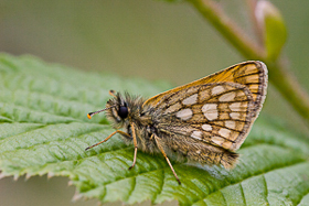
The body of the adult butterfly is comprised of 3 segments - head, thorax and abdomen. The eyes, antennae, proboscis and palpi are all positioned on the head. The legs and wings are attached to the thorax. The reproductive organs and spiracles are part of the abdomen. All of these features are discussed in detail below and the illustrations below provide an overview of the majority of these features.
 |
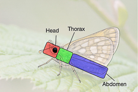 | 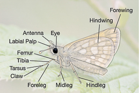 |
| Chequered Skipper (Carterocephalus palaemon) Image © Pete Eeles |
The head contains a pair of compound eyes, each made up of a large number of photoreceptor units known as ommatidia. Each ommatidium includes a lens (the front of which makes up a single facet at the surface of the eye), light-sensitive visual cells and also cells that separate the ommatidium from its neighbours. The image below shows a closeup of the head of a Pyralid moth, clearly showing the facets on the surface of the eye. A butterfly is able to build up a complete picture of its surroundings by synthesising an image from the individual inputs provided by each ommatidium.
 |
| Scanning electron microscope image of the head of a Pyralid moth Image © Dartmouth College |
The image above also highlights the coiled proboscis, which is made up of two concave parts which interlock, very much like a zip. The two parts are interlocked when the adult butterfly first emerges from the pupa, forming a tube through which nectar, minerals and moisture can be sucked up. The two parts of the proboscis can be separated for cleaning. Krenn (1997) provides a nice account of the joining of the two parts of the proboscis when the adult butterfly first emerges. Some butterflies, such as the Wood White (Leptidea sinapis) and Cryptic Wood White (Leptidea juvernica), also use their antennae as tactile (touch) instruments during courtship. In the case of these whites, the male and female face each other with wings closed and intermittently flash open their wings. At the same time, the male waves his proboscis and white-tipped antennae either side of the female's head. If the female is receptive to these signals, the female bends her abdomen toward the male and the pair mate.
| Cryptic Wood White (Leptidea juvernica) Courtship ritual Video © Pete Eeles |
The head also contains a pair of segmented antennae which act as sensors for a variety of purposes, including pheremone detection during mate location and when sensing the chemical properties of plants during feeding and ovipositing. The Monarch butterfly (Danaus plexippus) also uses time-compensated sun compass orientation during migration, which is supported by antennal clocks, as discussed in Froy (2003). Some butterflies, such as the Small Tortoiseshell (Aglais urticae) and Grayling (Hipparchia semele), use their antennae as tactile instruments during courtship. Butterfly antennae come in a variety of forms and are often used as an aid to classification. In general, butterfly antennae differ from those of moths in having a clubbed tip.
At the front of the head are a pair of labial palpi which serve a number of purposes. The first is to provide a level of protection to the eyes against debris such as dust and pollen grains, and they may also afford a level of protection to the proboscis. The palpi also act as tactile (touch) and olfactory (smell) sensors.
The thorax is the butterfly's engine room, containing the muscles that power the wings. The thorax is made up of three segments, each of which has a pair of legs attached to it. The second and third segments also have a pair of wings attached to them.
A butterfly has 3 pairs of legs - the forelegs, midlegs and hindlegs. In some species (such as those in the Nymphalidae family), the forelegs are reduced and no longer used for walking. Each leg has 3 main sections, the femur, tibia and tarsus, and the tarsus may end with a "claw". The tibia may also be decorated with one or more spines.
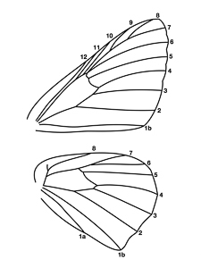
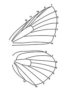
All butterflies have 2 pairs of wings - the forewings and hindwings. A network of rigid veins provides each wing with a structural framework, keeping it in shape. The venation (vein formation) is consistent between species and genera, and is used in butterfly classification. The venation of representative species is shown in the table below, with one representative for each family with the exception of Nymphalidae where the subfamilies of Danainae, Satyrinae and Nymphalinae are shown.
Please note: All line drawings are adapted from original drawings by and are © Timothy Freed, in Moths and Butterflies of Great Britain and Ireland, Volume 7, Issue 1 (Emmet & Heath, 1989).
 |  |
| Riodinidae Duke of Burgundy Hamearis | Lycaenidae Common Blue Polyommatus icarus |
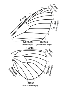
Various conventions exist for naming the veins and two are shown below. The first is used in this website.
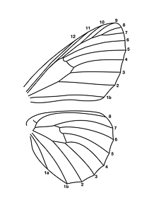 | 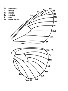 |
| Vein numbering | Vein numbering (alternative) |
The spaces between the veins and also various areas of the wings also have associated terminology and these are also shown in the two figures below.
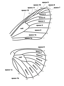 |  |
| Wing spaces | Wing regions |
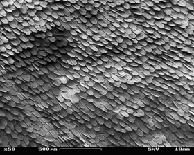
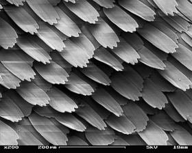
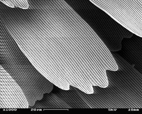
In most butterfly species, and all found in the British Isles, the transparent wing membranes are covered in overlapping scales, much like the tiles on a roof, as shown in the images below of a Peacock (Inachis io) wing. Each scale is a single cell and different cells vary considerably in shape - a feature that is used to distinguish certain species and subspecies. For example, a feature that distinguishes the various subspecies of Green-veined White (Pieris napi) is the shape of their androconial scales (and this type of scale is discussed below).
 |  |
| Peacock wing scales (x 50) | Peacock wing scales (x 250) |
 | 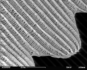 |
| Peacock wing scales (x 1000) | Peacock wing scales (x 5000) |
The colours produced by the scales are normally a result of the pigments that give each scale its colour. However, in some species, such as the Purple Emperor (Apatura iris), Small Copper (Lycaena phlaeas) and the spectacular Morpho butterflies, the apparent colour is actually the result of the nano-optical effects of reflection, refraction, diffraction and interference. Pantelic (2011) discusses the iridescence of the Purple Emperor (A. iris) and Lesser Purple Emperor (A. ilia), and Michielsen (2010) discusses how the green colouring of the Green Hairstreak (Callophrys rubi) is actually the result of light reflecting on crystals in the wing scales.
Other specialised scales include the androconial scales found on the forewings of the male in some species, such as within the costal fold of a Grizzled Skipper (Pyrgus malvae) or within the sex brand of a Silver-washed Fritillary (Argynnis paphia). These scales contain a pheremone at their base that is used during courtship to entice a female to mate.
The abdomen consists of 10 segments and is home to some key functions of the adult butterfly, namely the organs used in digestion, reproduction and breathing and the latter two are discussed in more detail here. The 10 segments of the abdomen are linked by tissues that allow the abdomen to bend (sometimes at quite acute angles) during copulation, egg laying or when (in the case of some Pieridae) a female is rejecting the advances of a potential suitor by pointing her abdomen upwards.
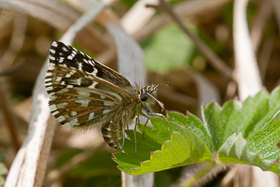 | 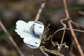 |
| Grizzled Skipper (Pyrgus malvae) Female ovipositing Image © Pete Eeles | Orange-tip (Anthocharis cardamines) Female rejecting male Image © Pete Eeles |
The end of the abdomen is home to the genitalia which are unique to each species, with the male and female organs of the same species acting as a matching "key and lock". Differences in genitalia are used in extreme circumstances to differentiate one species from another. For example, the Wood White (L. sinapis) and Réal's Wood White (L. reali) were originally identified as separate species based on differences in the genitalia, Réal's Wood White subsequently split into two species based on DNA analysis. The female butterfly is equipped with an ovipositor for egg laying which, in some species, can be quite robust. The Small, Essex and Lulworth Skippers (Thymelicus sylvestris, T. lineola and T. acteon), for example, are able to tease apart grass sheaths with their ovipositors before inserting a small number of eggs into the sheath.
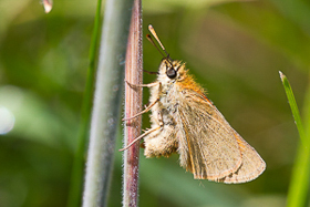 |
| Small Skipper (Thymelicus sylvestris) Using ovipositor to lay within a grass sheath Image © Pete Eeles |
The imago, larva and pupa all breathe through tiny holes in the sides of the abdomen, known as spiracles.
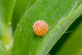
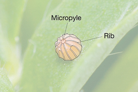
Butterfly eggs vary greatly in terms of size, shape and colour. However, the shape of an egg is often a good indication of which family or genus it belongs to, and this knowledge is often used in butterfly classification. Eggs may be adorned with structural ribs as shown in the figure below. A common characteristic of all eggs, however, is that they all have a micropyle, which is a small opening in the top of the egg through which sperm passes when the egg is fertilised.
 |  |
| Image © Pete Eeles | Image © Pete Eeles |
| Dingy Skipper (Erynnis tages) Image © Pete Eeles |
The larva is the only stage in which the butterfly grows and is sometimes viewed as little more than an eating machine. However, as the larva gets larger, it outgrows its skin and needs to moult in order to change into a new skin. Each period spent in a particular skin is known as an instar. The last skin change reveals the pupa.
| Wood White (Leptidea sinapis) Larva pupating (real time) Video © Pete Eeles |
As for all other stages, larvae are highly variable, even within different instars of the same species. For example, a newly-emerged first instar Purple Emperor (Apatura iris) larva doesn't have the "horns" that are characteristic of later instars. That being said, the larval features are sometimes used as an aid to classifying a given genus or species of butterfly.
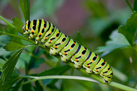
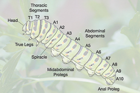
Each larva has 3 pairs of true legs, one pair on each thoracic segment. In addition, there are 4 pairs of "midabdominal" prolegs as well as a pair of anal prolegs at the rear end of the larva, all prolegs located on abdominal segments. As mentioned earlier, the larva also has a series of spiracles that allow it to breathe.
 |  |
| Image © Pete Eeles | Image © Pete Eeles |
| Swallowtail (Papilio machaon) Image © Pete Eeles |
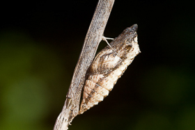
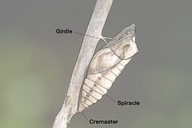
The pupa is the stage in which the butterfly transforms itself into the adult insect. Pupae are largely inactive but may wriggle (in some cases, quite violently) if they sense a potential threat. As with other butterfly stages, pupae come in all manner of shapes and sizes and are formed in a variety of positions and, again, these characteristics are sometimes used as an aid to classification. Most pupae are attached to a silk pad by the cremaster, found at the tail end of the pupa and typically adorned with a number of cremastral hooks, very much like the mechanism used in Velco.
 |  |
| Image © Pete Eeles | Image © Pete Eeles |
| Swallowtail (Papilio machaon) Image © Pete Eeles |
It is known that some butterfly pupae are capable of making sounds by moving different sections of the abdomen against each other. In the case of the Purple Hairstreak (Favonius quercus) it is thought that this mechanism is used to attract ants that subsequently clean and protect the pupa.
During the pupal stage, the larval structures are largely broken down while the adult structures of the adult butterfly are formed. The time-lapse video below shows the development of a Painted Lady (Vanessa cardui) butterfly within the pupal case, using an X-ray scanner. The shape of the adult butterfly can be made out by day 15; the eyes are at the bottom and its antennae, proboscis and legs are folded up along its front.
| Painted Lady (Vanessa cardui) Pupal development (time lapse) |
The emergence of the adult butterfly from its pupa is surely one of the most fascinating sights in the natural world. The adult butterfly emerges from the pupa (a process known as eclosion) and expands its wings by pumping haemolymph into the wing veins.
| Monarch (Danaus plexippus) Emerging from pupa (time lapse) |